Mon-fri: 9:00-19:00
Deutsch
German
Registrieren
Login
Dateien
Stock
Foto
Video
Audio
Vektor
Offer
Collections
Packages
Lightbox
Posten
Häufigste Downloads
Beste
Populärste
Neu
Kostenlos
Drucke und Produkte
Canvas Prints
Framed Prints
Prints and Posters
Metal Prints
Acrylic Prints
iPhone Cases
Galaxy Cases
Pillows
Duvet Covers
Shower Curtain
T-Shirts
Greeting Cards
Tote Bags
Kategorien
Portrait
Tiere
Video
Seiten Infos
Über uns
Support
Datenschutzerklärung
FAQ
Kontakt
Ihr Warenkorb ist leer.
Dateien
Stock
Foto
Video
Audio
Vektor
Offer
Collections
Packages
Lightbox
Posten
Häufigste Downloads
Beste
Populärste
Neu
Kostenlos
Drucke und Produkte
Canvas Prints
Framed Prints
Prints and Posters
Metal Prints
Acrylic Prints
iPhone Cases
Galaxy Cases
Pillows
Duvet Covers
Shower Curtain
T-Shirts
Greeting Cards
Tote Bags
Kategorien
Portrait
Tiere
Video
Seiten Infos
Über uns
Support
Datenschutzerklärung
FAQ
Kontakt
Suchen
Schlüsselwörter:
Creator ID:
Typ:
Foto
Illustration
Vektor
Video
Template
3d
Kategorien:
Animals
Buildings and Architecture
Business
Drinks
The Environment
States of Mind
Food
Graphic Resources
Hobbies and Leisure
Industry
Landscapes
Lifestyle
People
Plants and Flowers
Culture and Religion
Science
Social Issues
Sports
Technology
Transport
Travel
News
Entertainment
Sport News
Sprachen:
Brazilan Portuguese - Brazil
English - Canada
French - Canada
Spanish - Mexico
English - United States
English - Belgium
French - Belgium
Dutch - Belgium
Czech - Czech Republic
English - Cyprus
Danish - Danmark
German - Germany
English - Estonia
Spanish - Spain
French - France
French - Marocco
English - Greece
English - Ireland
Italian - Italia
English - Latvia
English - Lithuania
German - Luxembourg
English - Luxembourg
French - Luxembourg
English - Hungary
English - Malta
Dutch - Nederlands
Bokmal Norwegian - Norway
German - Austria
Polish - Poland
Portuguese - Portugal
English - Romania
German - Switzerland
English - Slovenia
Slovakian - Slovakia
French - Switzerland
English - Finland
Swedish- Sweden
Italian - Switzerland
English - United Kingdom
English - Bulgaria
English - Australia
Japanese - Japan
Korean - South Korea
English - New Zealand
Russian - Russia
English - Ukrain
English - Thailand
Ausrichtung:
Alle
Horizontal
Vertical
square
Farbe:
Model property release:
Alle
JA
NEIN
Laufzeit Video:
Alle
Up to 10 seconds
Up to 20 seconds
Longer than 30 seconds
Resultat
(0)
in die Webseite
Getty/iStock
Shutterstock
Fotolia
Depositphotos
123rf
Bigstockphoto
Pixabay
Adobe Stock
Relevance
Neu
Populärste
Häufigste Downloads
Undiscovered
12
24
36
48
96
Automatisch Seiten erstellen (Auto paging)
Menue
Brain anatomy, 3D illustration and composite MRI
CT scan showing right ventricular strain in a patient with pulmonary embolism, highlighting right ventricular enlargement and flattening of the interventricular septum
Human motor cortex, photomicrograph showing pyramidal neurons in cortical layers responsible for voluntary movement control and motor coordination.
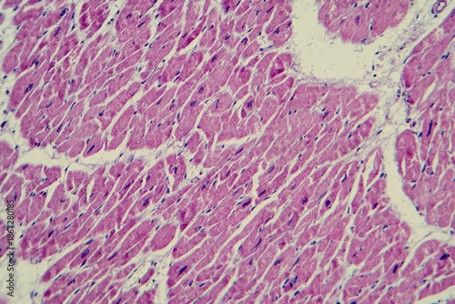
Cardiac hypertrophy, light micrograph
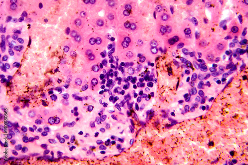
Red hepatization stage of lobar pneumonia, photomicrograph showing alveolar exudate with red cells, neutrophils, and fibrin strands
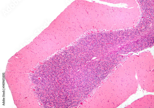
Human cerebellar cortex, photomicrograph showing Purkinje cells, granular cells, and molecular layer involved in motor coordination and balance.
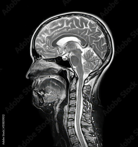
Axial T2-weighted brain MRI of a 40-year-old, showing midbrain, temporal lobes, and hippocampal region. No visible pathology, symmetrical structures, clear ventricles.
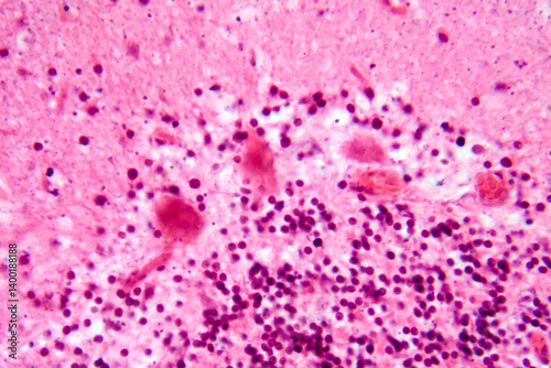
Coccidia in liver, photomicrograph showing intracellular parasitic protozoa causing hepatic coccidiosis with inflammation and hepatocellular damage.
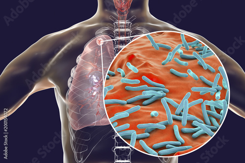
Secondary tuberculosis in lungs and close-up view of Mycobacterium tuberculosis bacteria, 3D illustration
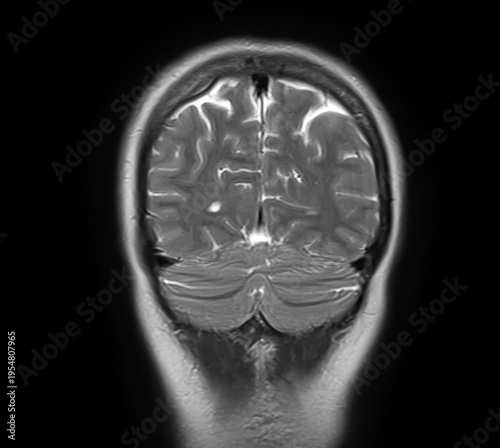
Normal brain MRI, coronal T2-weighted image showing cerebral hemispheres, cerebral cortex, cerebellum, cerebellar hemispheres, vermis and posterior fossa.
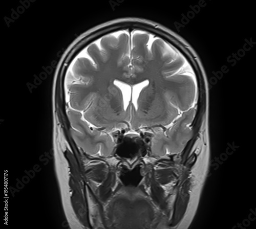
Normal brain MRI, coronal T2-weighted image showing cerebral hemispheres, lateral ventricles, cerebellum, cerebellar hemispheres, vermis and brainstem.
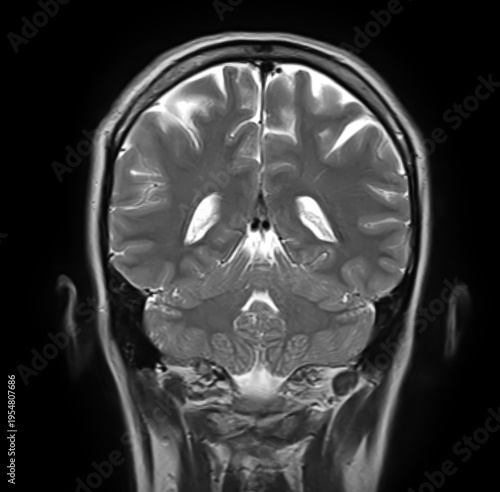
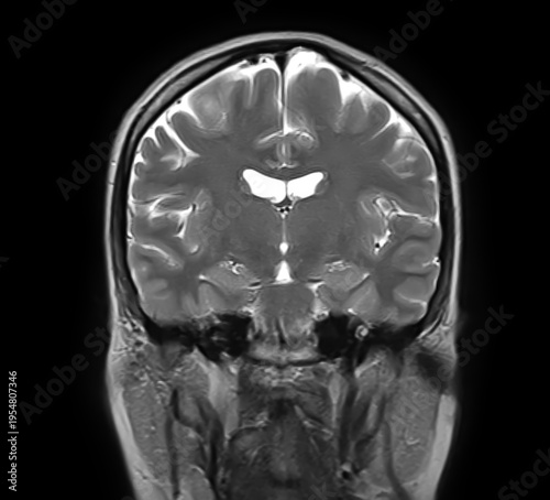
Normal brain MRI, coronal T2-weighted image showing cerebral hemispheres, cortex, lateral ventricles, mesial temporal lobes, hippocampi, brainstem and cerebellum.
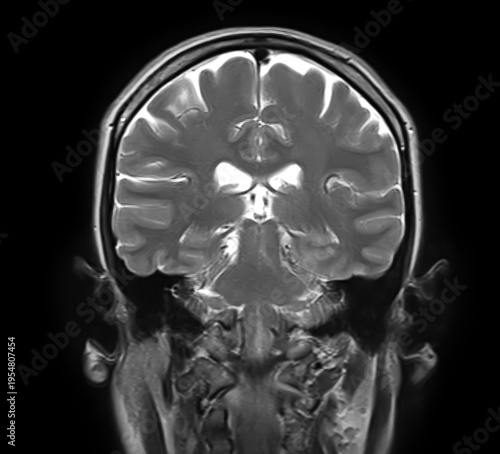
Normal brain MRI, coronal T2-weighted image showing cerebral hemispheres, cortex, lateral ventricles, basal ganglia, thalami, temporal lobes and mesial temporal structures.
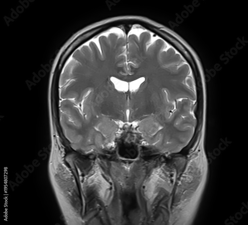
Normal brain MRI, coronal T2-weighted image showing cerebral hemispheres, cortex, lateral ventricles, basal ganglia, thalami, temporal lobes and sphenoid sinus.
Normal brain MRI, coronal T2-weighted image showing cerebral hemispheres, cortex, lateral ventricles, basal ganglia, thalami, temporal lobes and sphenoid sinus.
Normal brain MRI, coronal T2-weighted image showing cerebral hemispheres, cortex, lateral ventricles, basal ganglia, thalami, temporal lobes and sphenoid sinus.
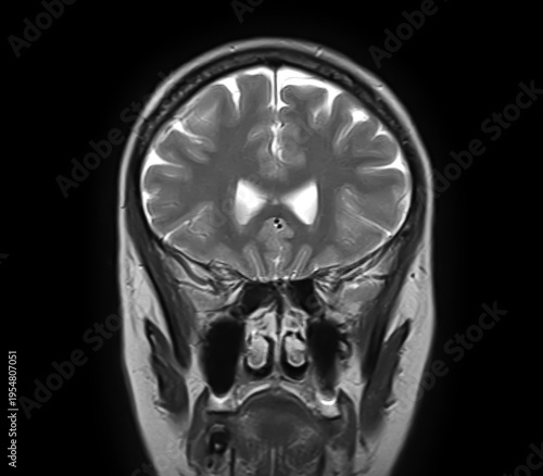
Normal brain MRI, coronal T2-weighted image showing cerebral hemispheres, cortex, lateral ventricles, ethmoid air cells, maxillary sinuses, nasal cavity and turbinates.
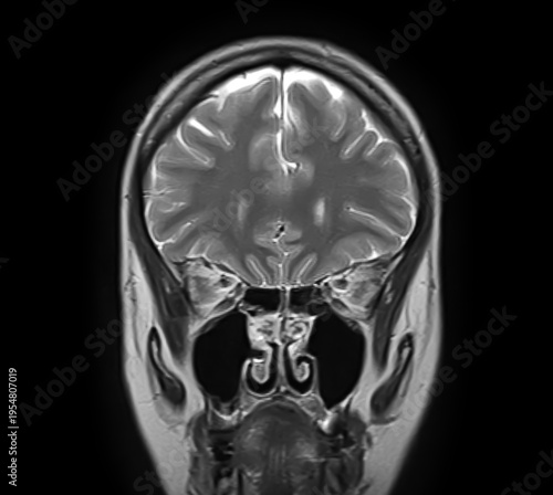
Normal brain MRI, coronal T2-weighted image showing frontal lobes, cerebral cortex, interhemispheric fissure, ethmoid air cells, maxillary sinuses, nasal cavity and turbinates.
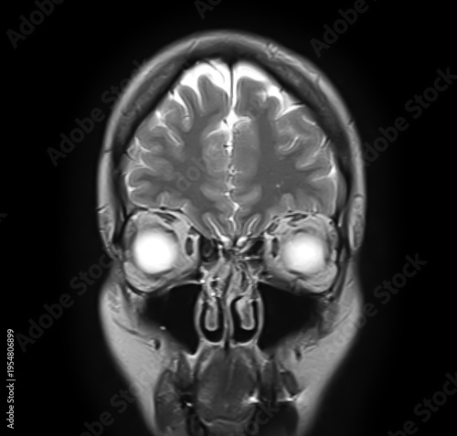
Normal brain MRI, coronal T2-weighted image showing frontal lobes, cerebral cortex, interhemispheric fissure, orbits, globes, ethmoid air cells, maxillary sinuses and nasal cavity.
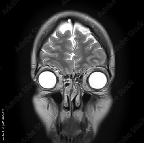
Normal brain MRI, coronal T2-weighted image showing frontal lobes, cerebral cortex, interhemispheric fissure, orbits, globes, ethmoid sinuses and nasal cavity.
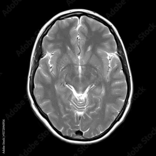
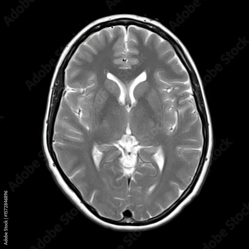
Axial T2-weighted brain MRI of a 40-year-old showing normal anatomy of lateral ventricles, thalamus, and basal ganglia without visible pathology.
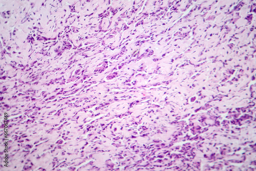
Osteosarcoma, a malignant bone tumor, light micrograph under the microscope
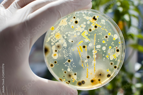
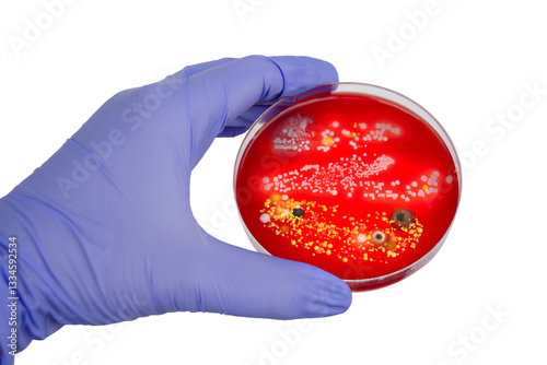
Researcher hand in glove holding Petri dish with colonies of different bacteria and molds on natural background. Biotechnology concept
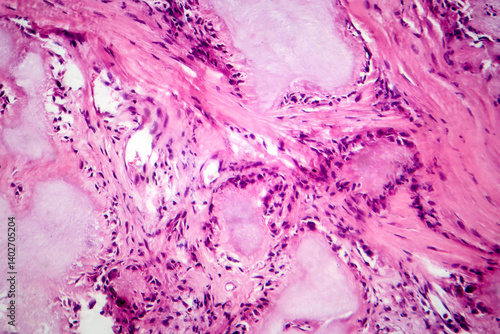
Gout tophus, photomicrograph showing amorphous, eosinophilic material with a foreign-body giant cell reaction and inflammation
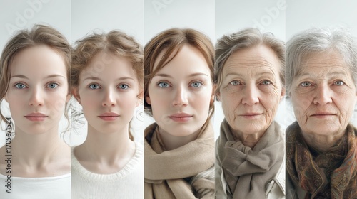
Photorealistic set of portraits of the same European person at different ages, from child to old, looking at camera on white background, generative ai illustration
Secondary tuberculosis in lungs, apical nodule, 3D illustration
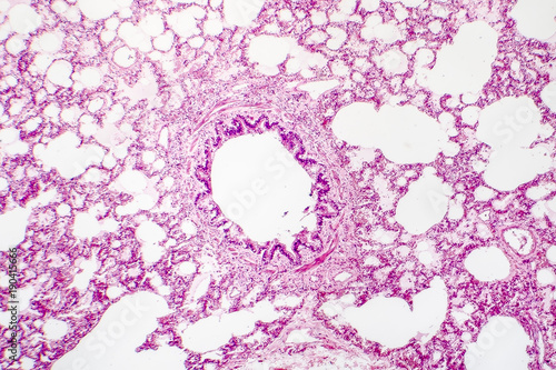
Cross-section of human lung tissue showing bronchiole and alveoli, histology, micrograph, photo under microscope
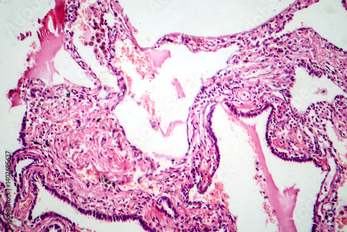
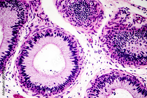
Histology of human epididymis tissue, micrograph. Photo under microscope.
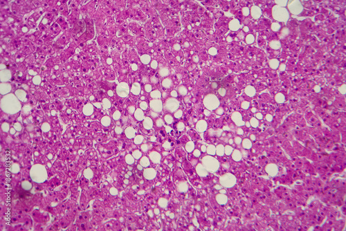
Hepatic steatosis, light micrograph
Bacterial and mold fungi colonies grown from indoor air on blood sheep agar
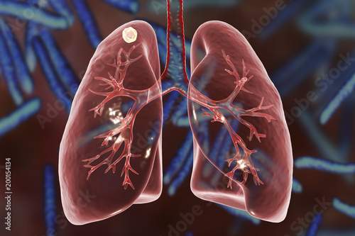
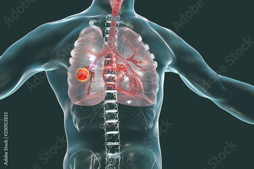
Lung cancer, medical concept, 3D illustration showing cancerous tumor inside human lung
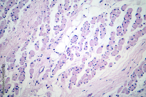
Skeletal muscle atrophy, photomicrograph showing decreased fiber size with increased spacing between them, reduced myofibrils, increased endomysial connective tissue with fatty infiltration
 Deutsch